
Tested Applications
| Positive WB detected in | Calyculin A treated HEK-293T cells, HEK-293 cells,Jurkat cells,Calyculin A treated PC-3 cells,TPA treated Jurkat cells |
| Positive IHC detected in | human breast cancer tissueNote: suggested antigen retrieval with TE buffer pH 9.0; (*) Alternatively, antigen retrieval may be performed with citrate buffer pH 6.0 |
Recommended dilution
| Application | Dilution |
|---|---|
| Western Blot (WB) | WB : 1:2000-1:10000 |
| Immunohistochemistry (IHC) | IHC : 1:100-1:400 |
| Sample-dependent, check data in validation data gallery | |
Published Applications
| WB | See 200 publications below |
| IHC | See 22 publications below |
| IF | See 1 publications below |
Product Information
66444-1-Ig targets Phospho-AKT (Ser473) inWB, IHC, IF, ELISA applications and shows reactivity with human,mouse samples.
| Tested Reactivity | human,mouse |
| Cited Reactivity | chicken, human, mouse, rat |
| Host / Isotype | Mouse / IgG1 |
| Class | Monoclonal |
| Type | Antibody |
| Immunogen | Peptide |
| Full Name | v-akt murine thymoma viral oncogene homolog 1 |
| Observed molecular weight | 60-62 kDa |
| GenBank accession number | NM_005163 |
| Gene symbol | AKT1 |
| Gene ID (NCBI) | 207 |
| Conjugate | Unconjugated |
| Form | Liquid |
| Purification Method | Protein A purification |
| Storage Buffer | PBS with 0.02% sodium azide and 50% glycerol pH 7.3. |
| Storage Conditions | Store at -20°C. Stable for one year after shipment. Aliquoting is unnecessary for-20oC storage. |
Background Information
1) What is AKT?
The serine/threonine kinase B AKT pathway (also known as the PI3K-Akt pathway) plays a vital role in the regulation of cellular processes, including cell proliferation, survival, and growth – processes that are essential for oncogenesis. Mutation of the regulator proteins PI3K and PTEN causes uncontrolled disruption within the PI3-kinase pathway, leading to the development of human cancers (1,2; see also AKT pathway poster for more details).
2) phospho-AKT and FAQs
A) What is the best way to normalize phosphorylated proteins analyzed by western blot?Normalize phospho-AKT and total AKT with your loading control (e.g. Actin, tubulin), then calculate the phospho/total ratio using these normalized values. Put more simply:1. Calculate the ratio of band intensities of a phospho-AKT band: the loading control.2. Calculate the ratio of band intensities of total AKT: loading control.3. Divide ratio obtained #1 by #2 to obtain a normalized value for comparison among different conditions. This procedure allows one to distinguish between a change in AKT expression and a change in the ratio of phospho-AKT.* If you are looking at the differences in a phospho-AKT expression resulting from an experimental condition (e.g., knockdown), you should also show the expression of total AKT to distinguish between a change in AKT expression (transcription/translation level) and a change in the AKT phosphorylation status.B) What is the observed molecular weight for AKT and phospho-AKT?Molecular Weight AKT – 56 kDaMolecular Weight phospho-AKT – 60 kDa (Figure 1)
Figure 1. WB: HEK-293 cell lysate was subjected to SDS PAGE followed by western blot with 60203-2-Ig (AKT antibody) and 66444-1-Ig (AKT-phospho-S473 antibody) at a dilution of 1:4000 incubated at room temperature for 1.5 hours.
C) Are there any special WB conditions to optimize staining of a phospho-AKT?Since this is a phosphorylated protein, 5% BSA is recommended over non-fat milk as a blocking agent.D) What are good positive and negative controls for a phospho-AKT?- Positive Control: HEK293 cells- Negative Control: Treatment with PI3K inhibitors (e.g. wortmannin)E) What species does this antibody react with?
Our internal testing has confirmed that it reacts with the human and mouse forms of phospho-AKT.Reactivity with the human form is also supported by the literature’s citations of this antibody.
References:
1. Perturbations of the AKT signaling pathway in human cancer.2. Targeting the PI3K-Akt pathway in human cancer: rationale and promise.
Protocols
| Product Specific Protocols | |
|---|---|
| WB protocol for Phospho-AKT (Ser473) antibody 66444-1-Ig | Download protocol |
| IHC protocol for Phospho-AKT (Ser473) antibody 66444-1-Ig | Download protocol |
| Standard Protocols | |
|---|---|
| Click here to view our Standard Protocols |
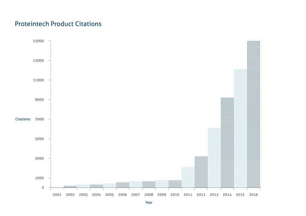
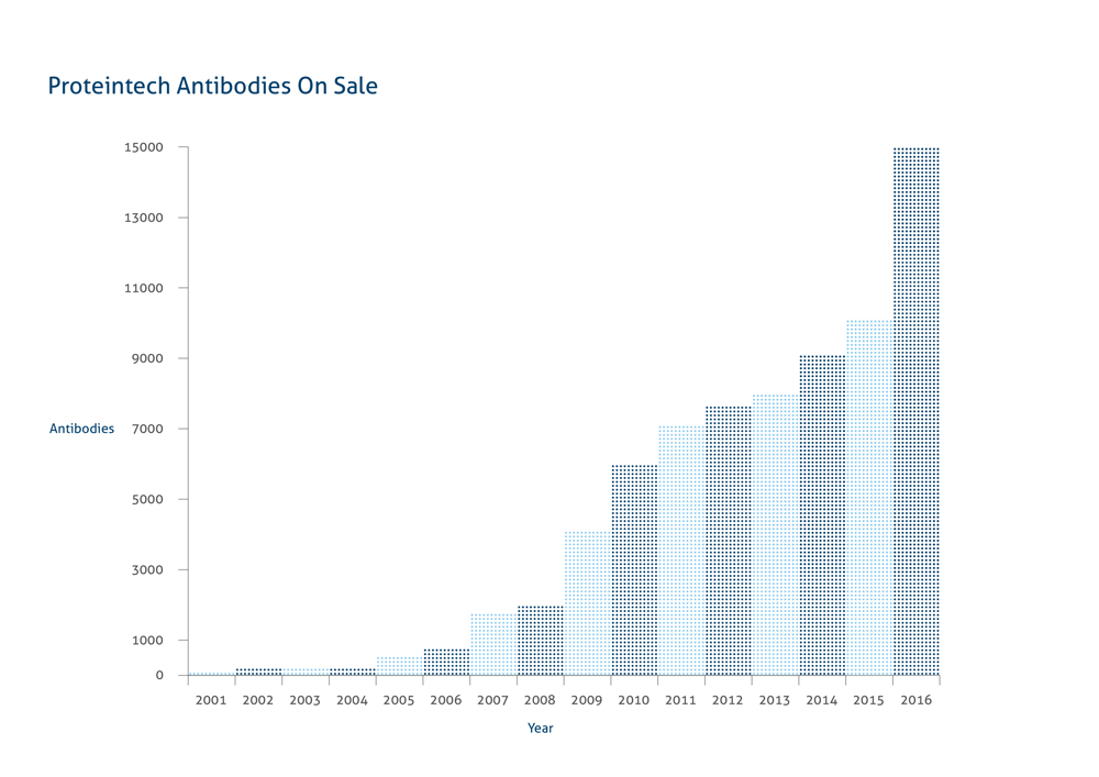
Proteintech是一家以研发、生产、销售抗体/重组蛋白及相关 产品为主要业务的生物技术公司。拥有严格的抗体生产、质检质控体系,产品涵盖整个生命科学领域。Proteintech作为全球最大的专业抗体生产 商之一,凭借一支锐意进取的技术团队和稳定的技术平台,已研发和生产出14000多种特种抗体和14000多种人源蛋白产品。2018年4月正式收 购首家人源细胞生产人类蛋白的Humanzyme公司,新增Humankine高活性人源蛋白产品系列。目前,Proteintech已建立全球市场销售网络, 美国、欧洲、中国和日本均有现货库存,全球24小时现货发售。

2001
Proteintech在美国创立

2003.8
使用Proteintech产品的第一篇文献发表

2004
在中国武汉成立中国分公司;第100个抗体成功生产

2001
Proteintech在美国创立

2003.8
使用Proteintech产品的第一篇文献发表

2004
在中国武汉成立中国分公司;第100个抗体成功生产

2006.4
第一个单克隆抗体上线销售

2007
在英国曼彻斯特成立欧洲分公司;使用Proteintech产品的发表文章达到50篇

2008
使用Proteintech产品的发表文章达到100篇

2001
Proteintech在美国创立

2003.8
使用Proteintech产品的第一篇文献发表

2004
在中国武汉成立中国分公司;第100个抗体成功生产

2006.4
第一个单克隆抗体上线销售

2007
在英国曼彻斯特成立欧洲分公司;使用Proteintech产品的发表文章达到50篇

2008
使用Proteintech产品的发表文章达到100篇

2010
使用Proteintech产品的发表文章达到1000篇

2012.3.3
第一个ELISA试剂盒上线销售;欧洲分公司获得ISO9001认证

2013
新实验室成立;使用Proteintech产品的发表文章达到5000篇

2001
Proteintech在美国创立

2003.8
使用Proteintech产品的第一篇文献发表

2004
在中国武汉成立中国分公司;第100个抗体成功生产

2006.4
第一个单克隆抗体上线销售

2007
在英国曼彻斯特成立欧洲分公司;使用Proteintech产品的发表文章达到50篇

2008
使用Proteintech产品的发表文章达到100篇

2010
使用Proteintech产品的发表文章达到1000篇

2012.3.3
第一个ELISA试剂盒上线销售;欧洲分公司获得ISO9001认证

2013
新实验室成立;使用Proteintech产品的发表文章达到5000篇

2014
获得SPF鼠房认证以及ISO9001以及ISO13485认证

2015.1
内部siRNA部门正式成立;使用Proteintech产品的文章达到10000篇

2018
Proteintech acquires Humanzyme, manufacturer of human-cell expressed proteins.

2001
Proteintech在美国创立

2003.8
使用Proteintech产品的第一篇文献发表

2004
在中国武汉成立中国分公司;第100个抗体成功生产

2006.4
第一个单克隆抗体上线销售

2007
在英国曼彻斯特成立欧洲分公司;使用Proteintech产品的发表文章达到50篇

2008
使用Proteintech产品的发表文章达到100篇

2010
使用Proteintech产品的发表文章达到1000篇

2012.3.3
第一个ELISA试剂盒上线销售;欧洲分公司获得ISO9001认证

2013
新实验室成立;使用Proteintech产品的发表文章达到5000篇

2014
获得SPF鼠房认证以及ISO9001以及ISO13485认证

2015.1
内部siRNA部门正式成立;使用Proteintech产品的文章达到10000篇

2018
Proteintech acquires Humanzyme, manufacturer of human-cell expressed proteins.
现在
Proteintech的产品已经达到12000多种,通过直销以及授权分销体系,产品覆盖五大洲55个国家。


